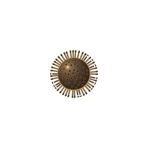
Influenza virus
Not alive in the usual sense: a shell of protein and fat carrying eight short strands of genetic instructions.
An influenza virus particle is usually a rough sphere about 80 to 120 nanometres across, some fifty times the width of the DNA helix. That is below the resolving power of an ordinary light microscope, which stops at about 200 nanometres, so flu viruses were first seen only with electron microscopes in the 1940s.
Inside a fatty envelope taken from the last cell it infected sit eight separate segments of RNA, holding the code for about a dozen proteins. The surface bristles with hundreds of spikes of two kinds: haemagglutinin, which latches onto cells lining your airways, and neuraminidase, which cuts newly made viruses free. Their varieties give strains names such as H1N1 or H3N2.
Because the genome comes in pieces, two different flu viruses infecting the same cell can swap segments. This reshuffling, together with error-prone copying, lets the virus change enough from year to year that flu vaccines have to be updated each season.
A virus has no metabolism and cannot reproduce on its own. It must enter a living cell and take over its machinery, and one infected cell can release thousands of new particles. Set against the E. coli bacterium, the next stop on this scale, a flu virus is roughly what a football is to a car.
- Size
- 1 × 10⁻⁷ m
- Typical diameter
- 80–120 nm
- Genome
- 8 RNA segments
- Surface spikes
- HA and NA proteins